Deep Learning for Distinguishing Morphological Features of Acute Promyelocytic Leukemia

Abstract
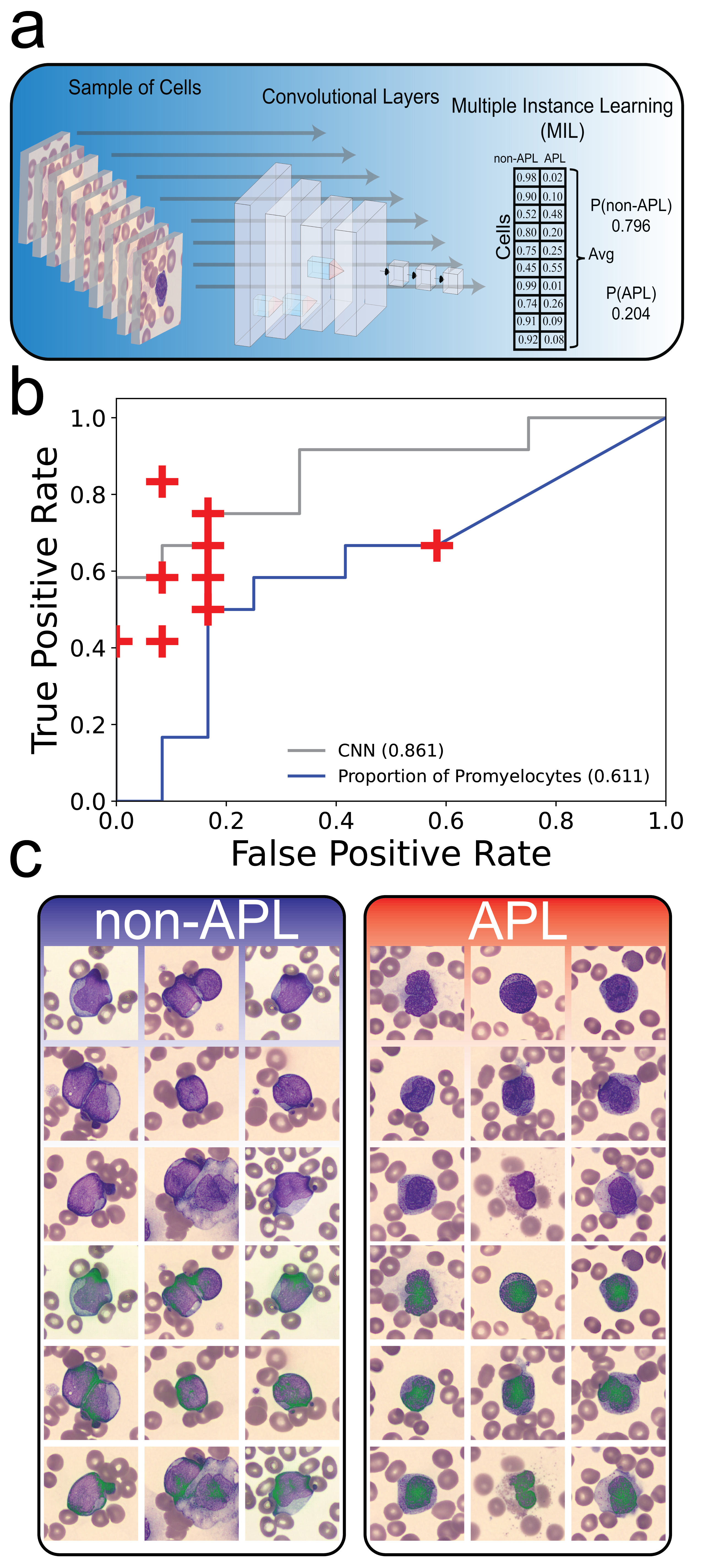
Background: Acute Promyelocytic Leukemia (APL) is a subtype of Acute Myeloid Leukemia (AML), classified by a translocation between chromosomes 15 and 17 [t(15;17)], that is notably distinguished clinically by a rapidly progressive and fatal course. Due to the acute nature of its presentation, prompt and accurate diagnosis is required to initiate appropriate therapy that can be curative. However, the gold standard genetic tests can take days to confirm a diagnosis and thus therapy is often initiated on high clinical suspicion based on both clinical presentation as well as direct visualization of the peripheral smear. While there are described cellular morphological features that distinguish APL, there is still considerable difficulty in diagnosing APL from direct visualization of a peripheral smear by a hematopathologist. We hypothesized that deep learning pattern recognition would have greater discriminatory power and consistency compared to humans to distinguish t(15;17) translocation positive APL from t(15;17) translocation negative AML. Methods: To best tackle the problem of diagnosing APL rapidly from a peripheral smear, study patients with APL and AML were identified via retrospective chart review from a list of confirmed FISH t(15;17)-positive (n = 34) and -negative (n = 72) patients presenting at The Johns Hopkins Hospital (JHH). Additional inclusion criteria included new disease diagnosis, no prior treatment, and availability of peripheral blood smear image uploaded to CellaVision. Patients were separated into a discovery cohort presenting prior to 1/2019 (APL, n = 22; AML, n=60) and a validation cohort presenting on or after 1/2019 (APL, n = 12; AML, n = 12). A multiple-instance deep learning model employing convolutional layers at the per-cell level (Figure 1A) was trained on the discovery cohort and then tested on the independent prospective validation cohort to assess generalizability of the model. Results: When compared to 10 academic clinicians (denoted with red +) who consisted of leukemia-treating hematologists, oncologists, and hematopathologists, the deep learning model was equivalent or outperformed 9/10 readers (Figure 1B) with an AUC of 0.861. We further looked at the performance of using proportion of promyelocytes (per CellaVision classification) as a biomarker of APL which had an AUC of 0.611. Finally, we applied integrated gradients, a method by which to extract per-pixel importance to the classification probability to identify and understand the morphological features the model was learning and using to distinguish APL (Figure 1C). We noted that the appearance of the chromatin in the non-APL leukemias was more dispersed and focused at the edge of the cell whereas in APL, the chromatin was more condensed and focused at the center of the cell. These morphological features, taught to us by the model, have not been previously reported in the literature as being useful for distinguishing APL from non-APL. Conclusion: Our work presents a deep learning model capable of rapid and accurate diagnosis of APL from universally available peripheral smears. In addition, explainable artificial intelligence is provided for biological insights to facilitate clinical management and reveal morphological concepts previously unappreciated in APL. The deep learning framework we have delineated is applicable to any diagnostic pipeline that can leverage a peripheral blood smear, potentially allowing for efficient diagnosis and early treatment of disease.
Presentation Video
Figures